
Sesamoid bone
| Sesamoid bone | |
|---|---|
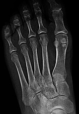
 Sesamoid bones at the distal end of the first metatarsal bone of the foot. | |
| Details | |
| Identifiers | |
| Latin |
|
| MeSH | D012716 |
| TA98 | A02.0.00.016 |
| TA2 | 374 |
| FMA | 32672 |
| Anatomical terms of bone | |
In anatomy, a sesamoid bone (/ˈsɛsəmɔɪd/)[1][2] is a bone embedded within a tendon or a muscle.[3] Its name is derived from the Greek word for 'sesame seed', indicating the small size of most sesamoids. Often, these bones form in response to strain,[4] or can be present as a normal variant. The patella is the largest sesamoid bone in the body. Sesamoids act like pulleys, providing a smooth surface for tendons to slide over, increasing the tendon's ability to transmit muscular forces.[3]
Structure

Sesamoid bones can be found on joints throughout the human body, including:
- In the knee—the patella (within the quadriceps tendon). This is the largest sesamoid bone.[4]
- In the flexor pollicis brevis). There is also commonly a sesamoid bone in distal portions of the second metacarpal bone and fifth metacarpal bone.[7]
- In the ossify in children ages 9–12.[9]
- In the metatarsal bone.
Common variants

- One or both of the sesamoid bones under the first
- The condyle of the femur. It is a variant of normal anatomy and present in humans in 10% to 30% of individuals. The fabella can also be mutipartite or bipartite.[13]
- The cyamella is a small sesamoid bone embedded in the tendon of the popliteus muscle. It is a variant of normal anatomy. It is rarely seen in humans, but has been described more often in other primates and certain other animals.[14]
-
![Lateral view.[11]](//upload.wikimedia.org/wikipedia/commons/thumb/7/7b/Accessory_and_sesamoid_bones_of_the_foot_-_lateral_projection.jpg/531px-Accessory_and_sesamoid_bones_of_the_foot_-_lateral_projection.jpg) Lateral view.[11]
Lateral view.[11] -
 Bipartite medial sesamoid bone under the first metatarsophalangeal joint of the great toe of the left foot of an adult woman.
![Lateral view.[11]](/File:Accessory_and_sesamoid_bones_of_the_foot_-_lateral_projection.jpg)

Clinical significance
- A common foot ailment in dancers is sesamoiditis (an inflammation of the sesamoid bones under the first metatarsophalangeal joint of the big toe). This is a form of tendinitis which results from the tendons surrounding the sesamoid becoming inflamed or irritated.[3]
- Sesamoid bones generally have a very limited blood supply, rendering them prone to avascular necrosis (bone death from lack of blood supply), which is very difficult to treat.[15]
Other animals
In
Although many carnivores have radial sesamoid bones,
Recently, the enlarged radial sesamoid bone of cotton rats has been studied.[23] Their enlarged radial sesamoid bone and that of the giant panda have a similar morphology and size relative to the rest of the hand.[23] The reason for this evolutionary change is still unknown; however, it may be to assist in grasping small objects and thin branches.[23]
Elephants have similarly enlarged sesamoid bones in both their forelimbs and hindlimbs, referred to as the prepollex and prehallux, respectively. These sesamoids function as "sixth toes", helping to distribute the animals' weight. In contrast to other sesamoids in elephants, which ossify at 3-7 years of age, the ossification of the prepollex and prehallux is delayed and is known to not have yet occurred in animals in excess of 20 years of age. The prehallux is further divided into two elements; the more proximal of these is fixed, whilst the more distal is mobile. Evidence of these "predigits" has also been found in certain fossil proboscideans.[24]
The forepaws of moles also possess a prepollex consisting of an enlarged, sickle-shaped sesamoid.
See also
Footnotes
- OED2nd edition, 1989 as /sεsəmɔɪd/
- ^ Entry "sesamoid" in Merriam-Webster Online Dictionary.
- ^ a b c "Sesamoid Injuries". aofas.org. Archived from the original on 2016-08-19. Retrieved 2014-12-06.
- ^ ISBN 978-0-07-337825-1.
- ^ Erica Chu; Donald Resnick (June 2014). "Sesamoid Bones: Normal and Abnormal". MRI Web Clinic. Retrieved 2017-11-04.
- PMID 26380010.
- ^ Erica Chu; Donald Resnick (June 2014). "Sesamoid Bones: Normal and Abnormal". MRI Web Clinic. Retrieved 2017-11-04.
- ISBN 978-0-12-746612-5.
- ISBN 978-0-07-337825-1.
- ISBN 978-0-12-746612-5.
- ^ a b Reference list for image is located at Commons:Template:Accessory and sesamoid bones of the foot - references.
- ^ Knipe, Henry. "Multipartite hallux sesamoid | Radiology Reference Article | Radiopaedia.org". radiopaedia.org.
- ^ Luijkx, Tim; Knipe, Henry. "Fabella". Radiopaedia. Retrieved 2015-09-18.
- S2CID 13339926.
- ^ "bunion, hammer toe, nail fungus, hallux rigidus". footankleinstitute.com.
- ^ PMID 17118063.
- ^ PMID 16387860.
- S2CID 16846874.
- PMID 11325067.
- PMID 11273049.
- S2CID 4302142.
- PMID 28096377.
- ^ S2CID 17239724.
- S2CID 206536505.
References
- Gray's Anatomy (1918) (Bartleby)
External links
 Media related to Sesamoid bone at Wikimedia Commons