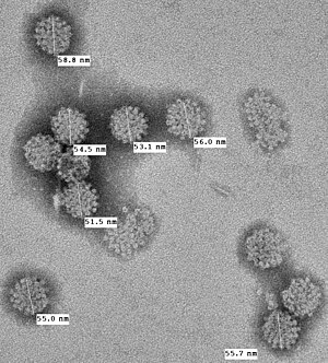
Merkel cell polyomavirus
| Human polyomavirus 5 | |
|---|---|
| Virus classification | |
| (unranked): | Virus |
| Realm: | Monodnaviria |
| Kingdom: | Shotokuvirae |
| Phylum: | Cossaviricota |
| Class: | Papovaviricetes |
| Order: | Sepolyvirales |
| Family: | Polyomaviridae |
| Genus: | Alphapolyomavirus |
| Species: | Human polyomavirus 5
|
Merkel cell polyomavirus (MCV or MCPyV) was first described in January 2008 in
Most MCV viruses found in MCC tumors, however, have at least two mutations that render the virus nontransmissible: 1) The virus is integrated into the host genome and 2) The viral T antigen has truncation mutations that leave the T antigen unable to initiate DNA replication needed to propagate the virus.[10]
Evidence that MCV is the cause for most MCC tumors comes from studies in which T antigen
Classification


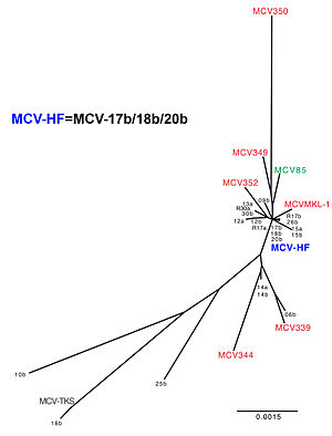
MCV is genetically most closely related to the
The prototype sequence of MCV has a 5387 base pair
MCV also expresses a microRNA (miRNA) known as MCV-miR-M1 from its late strand which bears perfect complementarity to LT and has been shown to negatively regulate LT expression .[17] In addition to its role in regulating MCV LT expression and DNA replication, MCV-miR-M1 has been shown to directly target and downregulate the expression of host cell immune related transcript SP100[18] and its role in the establishment of long-term persistent infection has been demonstrated in vitro.[19]
Viral cause for Merkel cell carcinoma
Merkel cell carcinoma is mainly seen in older individuals.
Eight of 10 Merkel cell carcinoma tumors initially tested were found to be infected with MCV.[1] In these tumors, the virus has integrated into the cancer cell genome and can no longer freely replicate. Recent studies from other laboratories have reproduced these findings: in one study 30 of 39 (77%) of Merkel cell tumors were MCV positive;[24] in another study, 45 of 53 (85%) Merkel cell tumors were positive.
Sequencing of the virus from Merkel cell cancers reveals that it generally has tumor-specific mutations that truncate the MCV T antigen. These mutations (which are not found in native virus obtained from nontumor sites) eliminate the T antigen helicase, preventing the integrated virus from replicating independently from the host cancer cell.
Prevention, diagnosis, and treatment
Persons who have Merkel cell carcinoma with this virus are not infectious to others and no infectious restrictions are warranted. The reasons for this are: 1) the virus in tumors is already mutated and no longer can be transmitted from tumors, and 2) most persons are already naturally exposed to this virus as children and young adults by other asymptomatic carriers.[citation needed]
Based on current data, prevention advice for MCC is similar to other skin cancers, such as avoiding sunburns and unnecessary sun exposure together with use of sun lotion. This may prevent mutations in the virus that increase risk for MCC among those already infected with MCV. Persons with immunosuppression (e.g., AIDS or organ transplant patients) are at higher risk for this cancer and may benefit from periodic skin examinations. Emergence of a painless lump that expands rapidly, especially among persons over age 50 or persons with immunosuppression, warrants examination by a physician. Biopsy of a Merkel cell tumor should readily provide a diagnosis and when caught early, has a good prognosis through standard treatment. At this time there are no vaccines or medications that can prevent MCV infection or prevent emergence of Merkel cell carcinoma.[citation needed]
Detection of the virus is still at a research phase and is generally not available as a clinical test. Detection of viral DNA is performed by PCR or by Southern blot. Caution is needed in interpreting results from PCR since it is prone to false-positive contamination and a substantial fraction of healthy skin samples may harbor low-level infection.[27] Sequencing of the viral genome may determine whether or not tumor-specific mutations are present.[citation needed]
Antibodies have been developed to stain for T antigen in tumor tissues [29] and appear to be specific for MCV-infected tumor cells.[30][31] Blood tests have also been developed[4][5] that show the majority of adults have been previously exposed to MCV and may continue to carry it as an asymptomatic infection.
Treatment guidelines do not differ for Merkel cell carcinoma infected with MCV or without MCV. A recent country-wide study from Finland suggests that MCV-positive tumors have a better prognosis than uninfected tumors[32] (although this has not been found in other studies[25]). If this is confirmed, routine detection of the virus may provide a future benefit for medical guidance. The virus itself is not known to be susceptible to current antiviral medications.
Recent studies reveal that the survivin oncoprotein is activated by MCV large T protein targeting the cellular retinoblastoma protein[33] and that survivin inhibitors can delay tumor progression in animal models. Clinical trials are now being organized to determine whether this has any benefit in humans. The importance of this finding is that a promising rational drug target was uncovered within four years of the initial discovery of the virus and that other new treatments might be rapidly developed now that the cause of the cancer is known. MCV is a target for cell-mediated immune responses, and so important research efforts are being focused on immunologic therapies that may benefit MCC patients.[citation needed]
Discovery and characterization
As a cause for Merkel cell carcinoma
While the original authors conservatively noted that it is "too early to tell" whether MCV is a cause of Merkel cell carcinoma, general scientific opinion now suggests that the virus causes most, but not all Merkel cell tumors. The virus is monoclonally integrated into the tumor when present, indicating that the proto-tumor cell was infected with the virus prior to its cancerous expansion. Mutations in the T antigen render the virus noninfectious, and therefore it is not a passenger virus that infected the tumor after the tumor had already started. Finally, the T antigen oncogene is expressed in all of the tumor cells and when it is inhibited ("knocked down" by
The reasons why 20% of Merkel cell carcinoma are negative for the virus remain completely unknown but speculations include the possibility that "Merkel cell carcinoma" is actually two or more closely related cancers, only one of which is infected with MCV. Misdiagnosis of this difficult cancer may also account for some of the negative results. Only a very small proportion of people infected with MCV develop the cancer. At this time no test for the presence of the virus is generally available, nor would patients be advised to change their treatment based on knowledge of MCV infection status. MCC patients can be enrolled in research studies, but these are not likely to directly benefit participants.[37] Reducing risk of UV exposure through sun screens is likely to reduce the risk of Merkel cell carcinoma as well as other skin cancers.
Moore has suggested that if his findings are confirmed, information about the virus could lead to a blood test or a vaccine that could improve the management of the disease or aid in prevention, much as the
Other associations
Possible associations with cervical carcinoma, cutaneous squamous cell carcinoma, Bowen's disease, basal cell skin carcinoma, extrapulmonary small cell carcinoma, and EGFR mutation-driven non-small cell lung cancer have been reported.[40][41][42][43][44][excessive citations]
References
- ^ PMID 18202256.
- ^ PMID 17686852.
- PMID 28174236.
- ^ PMID 19325891.
- ^ PMID 19499548.
- PMID 19239774.
- PMID 19239773.
- PMID 29238698.
- PMID 31549405.
- ^ PMID 18812503.
- PMID 20444890.
- ^ "Archived copy" (PDF). Archived from the original (PDF) on 2011-10-25. Retrieved 2012-07-17.
{{cite web}}: CS1 maint: archived copy as title (link) - ^ "Addgene: MCV-HF". www.addgene.com. Retrieved 20 April 2018.
- PMID 2998001.
- PMID 26218535.
- PMID 21841310.
- PMID 19046593.
- PMID 29777657.
- PMID 26218535.
- PMID 5009611.
- ^ S2CID 23833150.
- PMID 9625025.
- S2CID 11934339.
- ^ PMID 18593898.
- ^ PMID 18633441.
- PMID 21799863.
- ^ PMID 20542254.
- S2CID 35809601.
- PMID 19499546.
- S2CID 9819423.
- PMID 19609205.
- PMID 19535775.
- PMID 22572880.
- S2CID 29977325.
- S2CID 19709025.
- PMID 19078983.
- ^ "New Pathogen Discovery:Frequently Asked Questions". KSHV laboratory, molecular virology program, University of Pittsburgh Cancer Institute. Archived from the original on 2008-04-22. Retrieved 2008-04-13.
- ^ "Newly discovered virus linked to deadly skin cancer". University of Pittsburgh Medical Center News Bureau. 2007-01-17. Retrieved 10 July 2020.
- ^ Allison Gandey (2008-01-18). "newly discovered virus linked to neuroendocrine cancer of the skin". MedScape Medical News.
- PMID 22876976.
- PMID 20965777.
- PMID 19921198.
- PMID 24137462.
- PMID 24485957.
